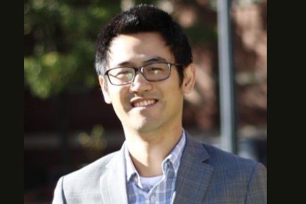
Ji Yi joined the Johns Hopkins Department of Biomedical Engineering as an assistant professor in September 2020. In this interview, Yi discusses his passion for science and engineering, his current research in designing and building microscopic tools to view biological structures, his goals as a new faculty member, and more.
What made you pursue a career in engineering?
Engineers not only settle on understanding the working mechanisms in nature, but also go on to translate the knowledge into making new things that do not exist. By that, engineering is a very rewarding and creative career that gives us the ability to visibly and directly make the world better.
Why did you choose Johns Hopkins BME? What are you looking forward to most?
Hopkins BME has held a special place in my heart. First, to state the obvious, it is the best BME department in the U.S.—and perhaps the world. I have known of Hopkins BME since my undergraduate studies in biomedical engineering at Tsinghua University. Several of my close friends joined the Hopkins BME graduate program and now are successful in their respective fields. There is also a personal connection because my brother and sister-in-law are also within JHU. It felt like a destiny call. What attracts me the most to Hopkins BME is the strong commitment to translational research to reshape and improve medicine by innovation. This is also what I am looking forward to most – the interaction with colleagues in an interdisciplinary and collaborative way.
Can you give a brief overview of your current research?
We are an imaging lab that designs and builds novel microscopic tools to visualize biological structures, process, and functions. Consider that any organism is constructed by building blocks, similar to LEGOs. 3D optical imaging allows us to virtually break down the structures and reveal the inner working principle of life in different length scales.
One area that we are actively working on is the vascular perfusion function. The vascular system delivers oxygen to support the local aerobic metabolism, and in turn, receives feedback from tissue to regulate the perfusion. The compromise of such interplay is implicated in a broad range of major diseases, including cancers, neurodegenerative diseases, and retinal vasculopathy. Our lab uses non-invasive imaging to quantify oxygen perfusion and metabolism from the 3D level down to the individual capillary level. The goal is to observe the interplay of the vascular perfusion to other tissue components in their native environment in 3D. Beyond vascular function, we are also developing large volume imaging and multimodal imaging to provide rich and complementary functional and structural information.
Have you ever experienced a “eureka moment?”
One moment occurred when I was a postdoc at Northwestern University developing visible light optical coherence tomography for retinal oximetry. We initially hypothesized that the auto-regulatory mechanism of the retinal circulation in the eye would compensate the systemic oxygen deficiency and maintain the oxygen metabolic rate (i.e. how fast oxygen was consumed measured by gas volume per unit time) by increasing blood flow. However, months and months of experimental repeats did not validate this hypothesis. Instead, it indicated an increase of oxygen consumption with systemic hypoxic challenges. We were deeply frustrated and puzzled until we realized that it could be the passive compensation to the deficiency from choroidal circulation beneath the retina. Then we worked with Professor Linsenmeier, who is an expert of oxygen in retina, and used his model to test this new theory. The curve aligned almost perfectly with our experiments, and everything clicked without missing dots. The moment that I saw the matching curve…EUREKA!
What do you consider your biggest research accomplishment so far?
We developed visible light optical coherence tomography (vis-OCT) for high-resolution imaging, reliable retinal oximetry, and spectroscopy. For the first time, we implemented this new technique in human retina. The sensitivity of spectroscopy can reach nanometer scale, way beyond the imaging resolution. We used non-invasive imaging to observe and quantify the interaction between two vascular systems on either side of the retina. The capillary-level oximetry and perfusion have strong clinical implications on various retinal vasculopathy, such as diabetic retinopathy, and age-related macular degeneration. My lab also pioneered mesoscopic oblique plane microscopy that allows volumetric imaging over a centimeter level field of view with isotropic 3D resolution. We have uniquely applied this technique in the ophthalmoscopy and are actively working on translating the technique in clinics.
What impact would you like your work to have?
I would like to contribute to biophotonics, and more broadly, the biomedical community, through our technical innovation. The new imaging capability in speed, resolution, imaging volume, and contrast can provide key information in answering questions that are otherwise difficult to study. Then, we would like our findings to help provide better solutions for biomedical challenges. Finally, we would like to translate the innovation in preclinical or clinical settings by offering sensitive and accurate imaging metrics, or seeing “invisibles.”
What are your goals for the future?
I am very excited about joining Hopkins BME, and my immediate goal is to develop a successful research program. Imaging can interact with many areas of research within the department and university. It will be a lot of fun working with colleagues in different disciplines to further fortify the leading research program as a whole. The ultimate goal is to use our knowledge to improve health care. After all, we are biomedical engineers.
Do you have any career advice to offer to current students?
Know where we are, where we come from, and where we are going. Be curious, and be willing to take some risks. And also ask tons of questions.
What do you enjoy doing outside the lab?
I am a foodie, a skier, a movie-lover, and a child-care giver for my two cute, little demons.

