
Hitting the target for successful heart ablation
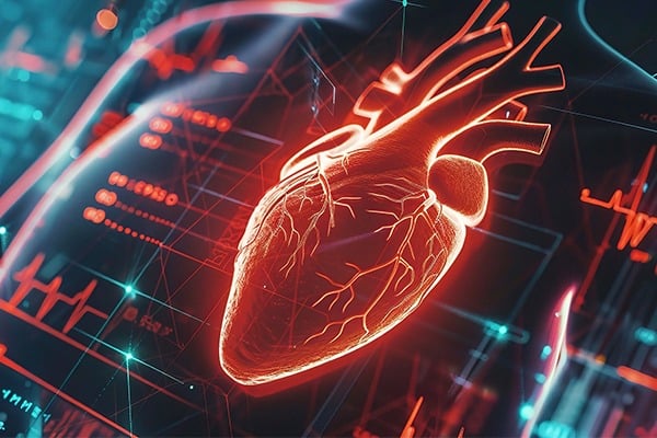
In a breakthrough for guiding the treatment of atrial fibrillation (AF), the most common form of heart arrhythmia affecting millions, Johns Hopkins researchers have for the first time developed digital twins of patients’ hearts that allow physicians to more precisely identify optimal targets for ablation — a procedure during which small patches of tissue causing the faulty rhythm are burned away.
This advance—based on a new understanding of how arrhythmias originate in diseased hearts and interact with burn lesions—promises to minimize lesions and reduce the need for repeated ablations, currently required for one-third of AF patients when the original procedure misses the problematic tissue regions.
“Ablation procedures, particularly in patients with persistent arrhythmia, can have very disappointing results because doctors don’t know where to burn, leaving the patient with unnecessary heart damage,” said study leader Natalia Trayanova, Murray B. Sachs Professor in the Department of Biomedical Engineering and director of the Alliance for Cardiovascular Diagnostic and Treatment Innovation at Johns Hopkins University. “By understanding how arrhythmias persist in each patient’s heart, we can inform clinicians how to best get rid of the problem tissue, while also preserving healthy heart tissue. With guidance from our models, doctors will be able to ablate only what they absolutely must to eliminate the arrhythmia.”
The team’s findings appear today in Nature Cardiovascular Research.
Trayanova’s lab has pioneered the development of personalized digital replicas of the heart based on the patient’s MRI scans and other data. In a previous study, her team demonstrated that the digital twin approach could successfully find ablation targets—regions where the heart’s electrical signals get disrupted and travel in a circular or rotational pattern instead of in a healthy, linear path.
In this more recent work, the team upped the ante: They aimed not only to identify the targets but also to optimize the ablation strategy for each patient’s unique tissue changes caused by AF.
Fibrosis, or scarring, of tissue in the two upper chambers of the heart (called “atria”) causes abnormal electrical activity and leads to arrhythmia. Ablation destroys the fibrotic tissue, interrupting the abnormal electrical signal. Even though ablation has been used for decades with variable outcomes, no prior studies explored the “ripple effect” of ablation to see how it alters the arrhythmia behavior elsewhere in the heart, said Trayanova. “What hasn’t been studied before is what happens to the electrical activity in the fibrotic tissue when you ablate a specific location in the heart. When you create damage in one area, the entire landscape for arrhythmias changes—what happens to the other areas? We used our digital twins to find out.”
The researchers performed 40 virtual ablations on 34 patients’ digital twin models. They found that when they ablated some target locations, other locations stopped being problematic (i.e. pro-arrhythmic) on their own. Based on this information, the researchers narrowed down which locations to target with ablation. Using this approach, they found that they could ultimately make the patient’s digital twin no longer able to sustain arrhythmia with 34% less burn lesions, preserving heart tissue.
Trayanova says the study represents a significant improvement over previous methods to predict ablation targets.
“What we demonstrate is a more comprehensive, non-invasive analysis of where to ablate using what we know about fibrosis and the fundamental mechanisms driving the arrhythmias. Before we could only tell where this rotational activity formed. With our new work, we can tell the clinician much more – which locations of rotational activity are important for sustaining atrial fibrillation and thus need to be ablated,” said Trayanova.
The work highlights a unique and extremely successful collaboration between Hopkins clinicians and engineers.
“Digital twins provide a unique opportunity to study physiology and personalize health care delivery in ways that more traditional therapeutic approaches cannot. Through close collaboration between engineers and physicians, we can create models, validate those models, design specific therapies tailored to that patient’s particular disease state, and then deliver that therapy clinically. It is a tremendously exciting process,” said David D. Spragg, associate professor in the Johns Hopkins University School of Medicine’s Division of Cardiology and co-author of the paper.
The team’s next steps are to implement the optimized digital twin prediction in a clinical trial and, eventually, usher the technology into clinical practice to make ablation faster and more effective for patients.
“First we treat the digital twin and then the clinicians will execute the predicted optimal ablation targets in the actual patient. They can achieve better results with more focused targets and significantly less ablation needed,” said Trayanova.
Additional Hopkins co-authors include Hugh Calkins, Kensuke Sakata, Ryan P. Bradley, Adityo Prakosa, Carolyna A. P. Yamamoto, Syed Yusuf Ali, Shane Loeffler, Brock M. Tice, Eugene G. Kholmovski, Ritu Yadav, Sunil Kumar Sinha, and Joseph E. Marine. Patrick M Boyle from the University of Washington also contributed to the work.