Database of Pediatric MR Images Aids Diagnosis, Treatment

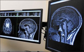
By building a “cloud database” of MR images collected from children with normal and abnormal brains, researchers aim to give physicians access to a Google-like search system that will improve the way pediatric brain disorders are diagnosed and treated.
The project is being developed by a team of engineers and radiologists at Johns Hopkins University School of Medicine, Baltimore, and is supported by a three-year, $600,000 grant from the National Institutes of Health, which is investing in Big Data to Knowledge (BD2K), an initiative to enable biomedical scientists to capitalize more fully on the Big Data being generated by those research communities.
“The project will allow doctors to search imaging records and find other annotated images in the database which ‘appear the same’ based on their anatomical phenotype,” said lead investigator Michael Miller, PhD, director of the Center for Imaging Science and a Hershel Seder Professor of Biomedical Engineering and Gilman Scholar at Johns Hopkins University. “This will allow them to compare their patient’s case to those that have already been logged and tagged as to etiology, disease and other clinical electronic medical records.”
Doctors will be also able to view and share brain MR images of children with diagnosed illnesses. By combining a structure-by-structure analysis of each child’s brain with an ontology of associated known pathology, the computer algorithm can “learn” what features are most closely related to different pathological conditions. As the computer gains more and more experience with expanding patients and studies, it will become better at recognizing subtle abnormalities.
The new data bank not only facilitates recognition and correct classification of pediatric brain disorders, but also the more objective image analysis allows identification of injury and disease that may go undetected by “eyeballing” MR images, said Thierry Huisman, MD, director of pediatric radiology at Johns Hopkins.
“Furthermore, recognition of distinct patterns of injury and the subsequent grouping of these children based upon their characteristic patterns of MRI findings allow recognition and identification of new diseases as well as reclassification of previously unclassified diseases,” Dr. Huisman said.
The pediatric data bank will search structural and functional imaging phenotype records to understand other cases that are similar and allow electronic medical record (EMR) annotation for training and correlation with rare and difficult cases, Dr. Miller said. With its machine learning capabilities, the data bank will also help with teaching and clustering to disease categories.
Currently, all radiology images are electronically archived in PACS, said Susumu Mori, PhD, professor and director of the Center for Brain Imaging Science in the Department of Radiology at John Hopkins. However, the description of the anatomy and the physician’s judgment process are largely unrecorded, save for a few sentences in the EMRs, Dr. Mori said.
“That expertise is shared through hands-on training, while a wealth of medical information remains in PACS and is not shared or reused, Dr. Mori said.
“For us, the images in PACS are somewhat akin to the blood samples in freezers,” Dr. Mori continued. “Theoretically, we can go back to the freezer and reanalyze the blood samples. Of course, we don’t do that because once the blood sample is analyzed and recorded into a chart, we can use the chart to share, search, analyze and build knowledge.”
By introducing a similar indexing technique, an image-based search will be possible. For example, certain image archives will allow text-based searches such as “retrieve all past cases with Batten disease.”
“We hope the image search will be a useful resource to support daily clinical routines,” Dr. Mori said.
There is a potentially broader impact as well, from using the resources as discovery tools. For example, in dementia cases, brain atrophy is often apparent in many patients. However, patterns of atrophy are very complex and their correlations with diagnosis and prognosis are often vague, Dr. Mori said.
“Even though humans have outstanding capability for feature extraction, a person is not as good as a computer at identifying pattern correlations on such a large-scale dataset,” Dr. Mori said. “Once all images in PACS are converted to indices, they become available for various relational analyses. This could deepen our understanding about the pathology and anatomical manifestations.”
The Power and Promise of Big Data Analytics
The project has been challenging in every area including the indexing accuracy, brain atlas inventory, cloud platform, database architecture and HIPAA compliance, Dr. Mori said. And ongoing challenges remain.
“These challenges are, however, precious experiences to help us deploy this technology in the clinical workflow,” Dr. Mori said. “The cloud platform is most useful if it is accepted in many other hospitals and enhances the information sharing and utilization. Sparsely indexed information, not dense, raw images, is the key for communicating imaging information.
“In addition, the platform will facilitate contributions from developers,” Dr. Mori continued. “Just like a travel website can provide services based on flight information from many air carriers, useful tools may emerge once the indexed data become widely available from many sources.”
What makes this project so important is that it is one of the first true manifestations of the power of “big data analytics,” said Jonathan Lewin, M.D., senior vice-president of Integrated Healthcare Delivery and the Martin Donner Professor and Chairman of the Department of Radiology and Radiological Science at Johns Hopkins.
“The ability to analyze thousands of examinations across multiple clinical conditions, combined with the advanced analysis tools developed by Drs. Mori and Miller, can serve as an ‘intelligent assistant’ for the radiologist, suggesting possible pathological conditions based on anatomic distortions and lesion characteristics in a manner resembling ‘CAD on steroids,’” said Dr. Lewin, who is also a professor of biomedical engineering, neurosurgery and oncology at Johns Hopkins. “Rather than the current level of CAD, in which the types of lesions being detected come from a very limited set of morphologies and densities, this new imaging intelligence and analytics approach can greatly broaden the power of the computer to assess expanding data sets of a growing range of pathologies.”
Beyond Johns Hopkins, the project could expand to aid other hospitals in pooling neuro data in common databases and searching and retrieving information, Dr. Miller said. The challenge would lie in data governance — in getting the partnering institutions to agree to participate and share their data in compliance with HIPAA regulations and with appropriate security, Dr. Lewin said.
Technically, expansion would be relatively simple, Dr. Lewin said.
“The beauty of creating a cloud-based architecture is that it is ‘scale-free,’ meaning the basic database can be expanded to include other patients, other institutions or even other countries without requiring any change in the basic design,” Dr. Lewin said. “Since the software learns a little bit from every brain scan it encounters, the more institutions that are added, the ‘smarter’ it gets, assuming that the information about underlying pathology is accurate.
“This way, other institutions can join the ‘cloud’ without having to reinvent anything or duplicate all of the effort and investment that went into its creation.”
— Felicia Dechter
