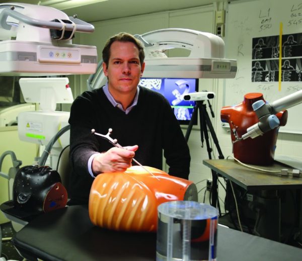
C-Arms Bring 3-D to the OR

Imaging has become an essential part of a surgeon’s toolkit in the past two decades, making operations on the most delicate parts of the body, such as the brain and the spine, safer and less invasive.
But there is major room for improvement, says Jeff Siewerdsen, the John C. Malone Professor in biomedical engineering and vice chair for clinical translation in that department. At the Carnegie Center for Surgical Innovation, which he established in 2016, Siewerdsen and his team are advancing imaging technologies that will make surgery more precise and improve patient safety.
A common technology in the operating theater are imaging devices called mobile C-arms: large, C-shaped devices with an X-ray source on one end and a detector on the other. The arm is positioned beside the OR table, providing high-resolution images of the patient, which surgeons and interventional radiologists use to navigate to their targets. High-tech C-arms developed in recent years allow 3-D imaging but still cannot visualize soft tissue.
“It’s difficult to approach a tumor and avoid normal tissue, nerves, and blood vessels,” Siewerdsen says. “Also, it’s hard to detect complications, like hemorrhage.”
He has led work to develop the first mobile C-arms for high-quality 3-D imaging of soft tissues. His team did this by converting a C-arm into a computed tomography system. Conventional CT scanners are large machines that spin quickly around a patient and combine x-ray projections taken from all the angles to reconstruct a 3-D image.
To impart this ability to a mobile C-arm, Siewerdsen began by motorizing it to rotate around the patient. They also changed the X-ray detector with a high-quality digital detector for cone beam CT—a method for 3-D imaging that uses a low-power fluoroscopic x-ray beam and only needs to rotate around the patient once.
Some resulting challenges: The low-power signal gives “noisy” images. And involuntary patient motion can cause distortions and streaks. To tackle these issues, Siewerdsen teamed with collaborators to develop advanced algorithms for high-quality image reconstruction that improves soft tissue image quality and reduces radiation dose. They are also developing “autofocus” methods that unravel motion-related distortions to restore image quality and avoid retakes. “These advances will allow a surgeon to more precisely approach the target, avoid normal tissue, and detect complications,” he says.
The research has been funded by grants from the National Institutes of Health and by research collaborations with Siemens and Medtronic. Siemens plans to roll out the first commercial system for high-quality 3-D C-arm imaging this year.
– Prachi Patel
