Skip to Content

Low gravity in space travel found to disrupt normal rhythm in heart muscle cells

September 24, 2024

Johns Hopkins scientists who arranged for 48 human bioengineered heart tissue samples to spend 30 days at the International Space Station report evidence that the low gravity conditions in space weakened the tissues and disrupted their normal rhythmic beats when compared to earth-bound samples from the same source.

The scientists said the heart tissues “really don’t fare well in space,” and over time, the tissues aboard the space station beat about half as strong as tissues from the same source kept on Earth.

The findings, they say, expand scientists’ knowledge of low gravity’s potential effects on astronauts’ survival and health during long space missions, and they may serve as models for studying heart muscle aging and therapeutics on Earth.

A report of the scientists’ analysis of the tissues will be published during the week of Sept. 23 in the Proceedings of the National Academy of Sciences.

Previous studies showed that some astronauts return to Earth from outer space with age-related conditions, including reduced heart muscle function and arrhythmias (irregular heartbeats), and that some, but not all, effects dissipate over time after their return.

But scientists have sought ways to study such effects at a cellular and molecular level in a bid to find ways to keep astronauts safe during long spaceflights, says Deok-Ho Kim, a professor of biomedical engineering and medicine at Johns Hopkins University. Kim led the project to send heart tissue to the space station.

To create the cardiac payload, scientist Jonathan Tsui, coaxed human induced pluripotent stem cells (iPSCs) to develop into heart muscle cells (cardiomyocytes). Tsui, who was a PhD student in Kim’s lab at the University of Washington, accompanied Kim as a postdoctoral fellow when Kim moved to Johns Hopkins University in 2019. They continued the space biology research at Johns Hopkins.

Tsui then placed the tissues in a bioengineered, miniaturized tissue chip that strings the tissues between two posts to collect data about how the tissues beat (contract). The cells’ 3D housing was designed to mimic the environment of an adult human heart in a chamber half the size of a cell phone.

To get the tissues aboard the SpaceX CRS-20 mission, which launched in March 2020 bound for the space station, Tsui says he had to hand carry the tissue chambers on a plane to Florida, and continue caring for the tissues for a month at the Kennedy Space Center. Tsui is now a scientist at Tenaya Therapeutics, a company focused on heart disease prevention and treatment.

Once the tissues were on the space station, the scientists received real-time data for 10 seconds every 30 minutes about the cells’ strength of contraction, known as twitch forces, and on any irregular beating patterns. Astronaut Jessica Meir changed the liquid nutrients surrounding the tissues once each week and preserved tissues at specific intervals for later gene readout and imaging analyses.

The research team kept a set of cardiac tissues developed the same way on Earth, housed in the same type of chamber, for comparison with the tissues in space.

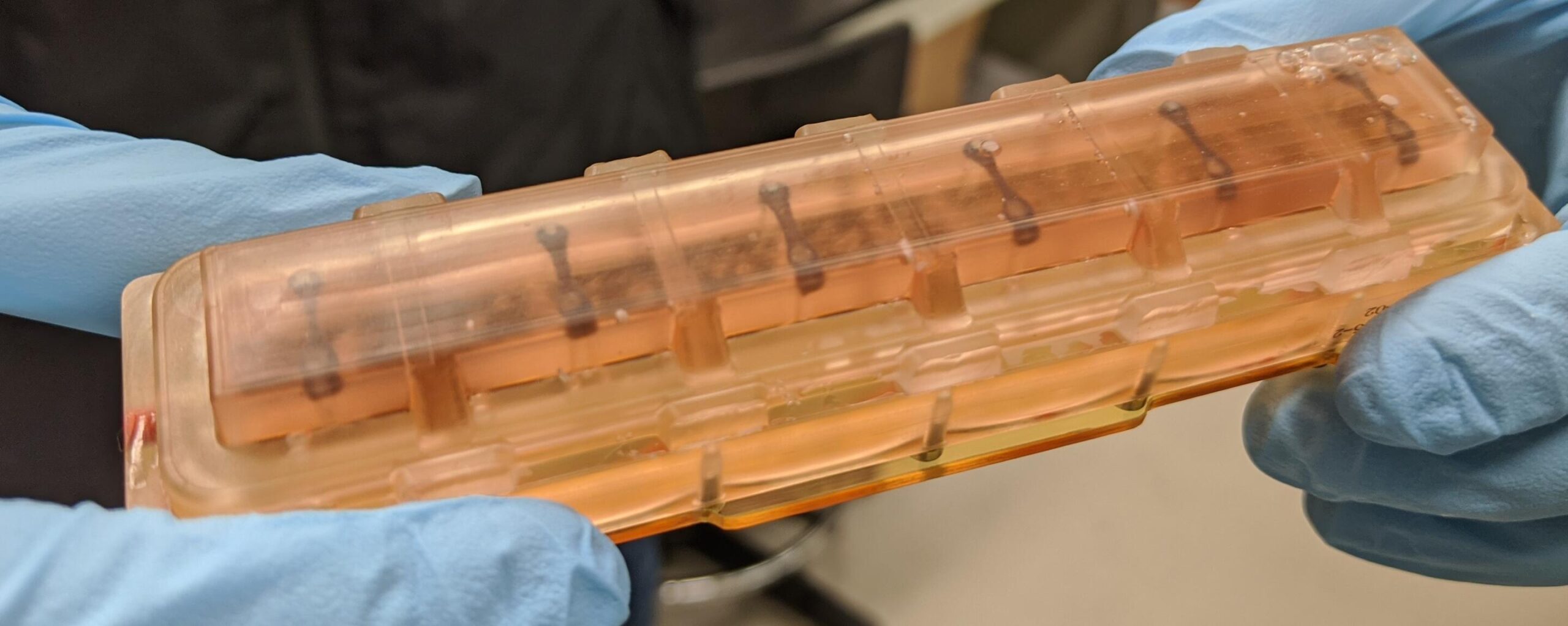
Heart tissues within one of the launch-ready chambers. PHOTO CREDIT: JONATHAN TSUI

When the tissue chambers returned to Earth, Tsui continued to maintain and collect data from the tissues.

“An incredible amount of cutting-edge technology in the areas of stem cell and tissue engineering, biosensors and bioelectronics, and microfabrication went into ensuring the viability of these tissues in space,” says Kim, whose team developed the tissue chip for this project and subsequent ones.

Devin Mair, a former PhD student in Kim’s lab and now a postdoctoral fellow at Johns Hopkins, then analyzed the tissues’ ability to contract.

In addition to losing strength, the heart muscle tissues in space developed irregular beating (arrhythmias)—disruptions that can cause a human heart to fail. Normally, the time between one beat of cardiac tissue and the next is about a second. This measure, in the tissues aboard the space station, grew to be nearly five times longer than those on Earth, although the time between beats returned nearly to normal when the tissues returned to Earth.

The scientists also found, in the tissues that went to space, that sarcomeres—the protein bundles in muscle cells that help them contract—became shorter and more disordered, a hallmark of human heart disease.

In addition, energy-producing mitochondria in the space-bound cells grew larger, rounder and lost the characteristic folds that help the cells use and produce energy.

Finally, Mair, Eun Hyun Ahn—an assistant research professor of biomedical engineering—and Zhipeng Dong, a Johns Hopkins graduate student, studied the gene readout in the tissues housed in space and on Earth. The tissues at the space station showed increased gene production involved in inflammation and oxidative damage, also hallmarks of heart disease.

“Many of these markers of oxidative damage and inflammation are consistently demonstrated in post flight checks of astronauts,” says Mair.

Kim’s lab sent a second batch of 3D engineered heart tissues to the space station in 2023 to screen for drugs that may protect the cells from the effects of low gravity. This study is ongoing, and according to the scientists, these same drugs may help people maintain heart function as they get older.

The scientists are continuing to improve their “tissue on a chip” system and are studying effects of radiation on heart tissues at the NASA Space Radiation Laboratory. The space station is in low Earth orbit, where the planet’s magnetic field shields occupants from most of the effects of space radiation.

Kim is a co-founder, scientific advisory board member and equity holder of Curi Bio, which develops bioengineered tissue platforms for drug development. Ahn is Kim’s spouse and serves as a co-investigator or principal investigator of NIH grants: UG3EB028094, UH3TR003519 and R21CA220111.

Funding for the research was provided by the National Institutes of Health (UG3EB028094, UH3TR003519, UH3TR003271, R01HL164936, R01HL156947, R21CA220111).

Other researchers who contributed to the study are Jeffrey Chen from Johns Hopkins, Ty Higashi, Alec Smith and Nathan Sniadecki from the University of Washington, Paul Koenig and Stefanie Countryman from the University of Colorado Boulder, and Peter Lee from Brown University.

Category: Research
Associated Faculty: Deok-Ho Kim

Read the Johns Hopkins University privacy statement here.

Accept