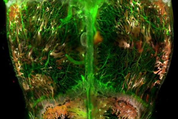
Johns Hopkins Medicine scientists have used glowing chemicals and other techniques to create a 3D map of the blood vessels and self-renewing “stem” cells that line and penetrate a mouse skull. The map provides precise locations of blood vessels and stem cells that scientists could eventually use to repair wounds and generate new bone and tissue in the skull.
“We need to see what’s happening inside the skull, including the relative locations of blood vessels and cells and how their organization changes during injury and over time,” says Warren Grayson, professor of biomedical engineering and director of the Laboratory for Craniofacial and Orthopaedic Tissue Engineering at the Johns Hopkins University School of Medicine. His lab focuses on developing biomaterials and transplanting stem cells into the skull to re-create missing bone tissue.
Other scientists have provided maps of small portions of blood vessels and stem cells in the mouse skull. “However, a larger picture of the skull gives us a better understanding of the entire vasculature and distribution of different stem cell types,” says Alexandra Rindone, graduate student at the Johns Hopkins University School of Medicine and first author of the paper.

