Virtual 3-D heart model guides defibrillator placement in children

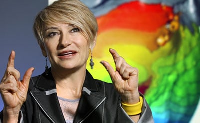
By joining MRI images with sophisticated computer analysis, lead investigator Natalia Trayanova, Ph.D., Johns Hopkins Murray B. Sachs Professor of Biomedical Engineering and co-investigator Jane Crosson, M.D. created a virtual 3-D heart model which analyzes a child’s anatomy and pinpoints the best location to implant a defibrillator device.
“Pediatric cardiologists have long sought a way to optimize device placement in this group of cardiac patients, and we believe our model does just that,” says Dr. Natalia Trayanova. “It is a critical first step toward bringing computational analysis to the pediatric cardiology clinic.”
If further studies show the model has value in patients, it could spare many children with heart disease from repeat procedures that are sometimes needed to re-position the device, says co-investigator Dr. Jane Crosson, a pediatric cardiologist and arrhythmia specialist at the Johns Hopkins Children’s Center. “It’s like having a virtual electrophysiology lab where we can predict best outcomes before we even touch the patient.”
A description of the team’s work, published ahead of print, can be found in The Journal of Physiology.
Read the Full Story: Johns Hopkins Medicine Press Release
