BME research lab receives funding and support to vastly improve CT scans

The CT scan machine — with a price tag of $1 million or more — is a marvel of modern medical diagnostic equipment. But even the best of them has some serious limitations.
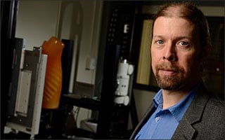
Assistant Professor J. Webster Stayman and his group in the Department of Biomedical Engineering’s Advanced Imaging Algorithms and Instrumentation (AIAI) Lab hope to improve on today’s CT scan devices through a four-year, $2.6 million grant from the National Institutes of Health. Their goal: further lower the amount of radiation patients receive during scans while maintaining or improving imaging performance.
Current CT scanners are limited in their ability to customize scans to the patient. Typically, technicians use basic information about a patient (e.g., a 6-foot, 180-pound male) to make basic adjustments to the X-ray exposure and energy. Higher exposures mean better image resolution but potentially can cause long-term harm to the patient. However, not all 6-foot, 180-pound men have the same body shape and density. So what’s optimal for one may not be for another.
Stayman and his team want to devise a way for the CT machine to automatically adjust radiation levels by doing a quick snapshot of the patient so it senses the body structure and tailors the exposure accordingly.
To customize the scan to the patient, Stayman and his colleagues are introducing dynamic X-ray beam-shaping hardware that directs the rays where they are needed most for high-quality imaging. The advances will mean modifications of both the hardware and software components of a CT scanner. These investigations will be made possible in part because one of the project’s participants is Philips Healthcare, one of the world leaders in the CT scan machine market. Philips will provide CT scan equipment for Stayman’s group to work with.
Besides making CT scans patient-specific, Stayman wants to make them more task-specific — detecting abnormalities in a specific organ, for example. But again, internal organs vary between people in shape and size, too. Accordingly, the advanced diagnostic device Stayman hopes to develop could customize its X-ray exposure — as well as the shape of the tomography beam it emits — based on the individual’s unique attributes and the body part being scanned.
Low-dosage CT scanners do exist, but keeping exposures to an absolute minimum is always a goal. And with better control of the tomography itself, physicians will literally have a clearer picture of the condition they’re treating.
— Michael Blumfield
